Image-based analysis in screening and diagnostics
Counting cells, measuring colonies, tracking organoids over time, classifying tissue sections – image analysis becomes the bottleneck in screening and diagnostics as soon as the number of images exceeds manual capacity. We build the models and pipelines that remove that bottleneck.
The situation
Automated microscopy produces more images than a laboratory can evaluate. Manual evaluation is not only slow but poorly reproducible: two analysts reach different counts on the same images.
Then there are technical confounders that cannot be ignored: illumination differences between plates, focus drift, edge effects in wells, batch differences in staining, touching and overlapping objects. A model that works on one plate and fails on the next moves the problem rather than solving it.
Typical data basis
Images from high-content screening, microscopy stacks, whole-slide preparations. Plus, and this is the limiting factor, annotations: the effort of labelling training examples is the real cost driver in practice, not compute time.
Why such initiatives fail
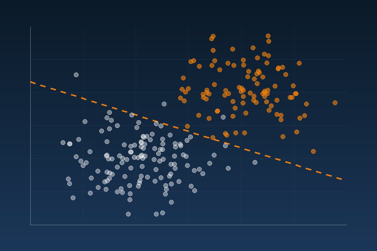
The model learns the plate instead of the biology. Where controls always sit in the same wells, or treatment groups are processed plate by plate, plate provenance is the strongest signal in the image. The model uses it – and fails as soon as the layout changes.
Splitting at image level instead of sample level. If tiles from the same preparation land in both training and test sets, the metric is massively overstated. The split has to happen at the level at which the decision will later be made: sample, patient, batch.
Annotation quality goes unmeasured. A model cannot become better than its reference. Without measured agreement between several annotators, nobody knows where the achievable ceiling lies – or whether 92% is a good result or a poor one.
Approach
The measurement first, the model second. What exactly is to be counted, measured or classified, and how accurate does the number have to be for a decision to rest on it? That question determines the architecture, not the other way round.
Segmentation before classification. Separating individual objects cleanly is the harder part with biological images. For cells and nuclei we use established specialised models such as Cellpose and StarDist; for large preparations, tiled processing with overlap handling.
Reduce annotation effort. Pre-trained models provide feature representations that transfer to a new question with only a few labelled examples. That shifts effort from labelling to analysis.
Test robustness rather than hoping for it. Evaluation broken out by plate, batch and site. We consider a model usable only when it works on images it has not seen in training.
Annotation with clinical expertise
Annotation quality caps model quality – and with clinical and histological images that annotation cannot come from student assistants. We work with a specialised annotation partner who annotates clinical data together with physicians.
That solves two problems at once. First, the reference is scientifically defensible rather than merely plausible-looking. Second, with several clinical annotators the agreement between them can be measured – which is what establishes the achievable ceiling for any model in the first place. Without that figure, every model metric lacks a yardstick.
What you get
Quantified measurements per object rather than one classification per image, a quality report that surfaces confounders, and a reproducible pipeline with a versioned model state – output in a form specialist teams can use without image-processing expertise.
Where our experience comes from
Image analysis is not a new discipline for us. We have built segmentation and detection models in projects where the same problems arise – small objects in large images, uneven illumination, overlapping structures:
- Pixel-accurate crack segmentation with a 97% true positive rate
- Object detection in orthophotos with over 99% detection rate
- Detection of bark beetle infestation in aerial imagery with Fast R-CNN
- Crop damage detection from drone imagery
The domain differs; the methodological task does not: turning a large image into reliably countable objects.
Last updated: 4 August 2026